Lameness Exams
Lameness is a generalized term for an abnormal gait in a horse and can occur for a wide variety of reasons. Determining the cause of a lameness is not always straightforward, and a lameness exam may involve a variety of diagnostic steps and treatment options.
A lameness exam consists of multiple parts, including both at rest and dynamic evaluations.
In a dynamic exam, your veterinarian will evaluate your horse in motion under a variety of conditions. This could include at a walk or jog in-hand on multiple types of surfaces (soft footing vs. hard ground), in straight lines and/or on a lunge line.
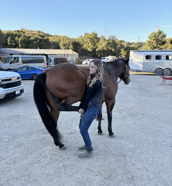
Flexion tests are a part of a dynamic exam where your veterinarian will flex and hold different portions of the lower limbs to try to accentuate a lameness and narrow down where the lameness is stemming from.
Local anesthesia or “blocks” may be utilized to further narrow down the location of a lameness. By strategically anesthetizing different nerves in the lower limb in a particular order until the lameness is improved, blocking is a common diagnostic procedure to pinpoint where a lameness may be coming from, may be used to rule out certain areas of concern, or where further diagnostics may be needed.
Further diagnostics in the field for lameness can include radiographs and ultrasound. These tools are most helpful to be utilized when a lameness has been localized to a specific area of concern or suspicion.
Once a cause for the lameness has been identified, your veterinarian can work with you to develop an appropriate treatment plan for your horse.
Certain lamenesses can be incredibly hard to diagnose. They may not block out using local anesthesia, may not have changes on radiograph or ultrasound, may be extremely subtle, or may involve multiple limbs. With difficult lameness cases, referral to a hospital for additional diagnostics including nuclear scintigraphy (bone scan), MRI, PET scan, or CT may be warranted.
